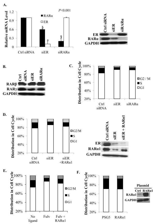
Figure 4.
Role of RARα in mediating the hormone-independent effect of ER on basal level cell cycling. (A) Effect of knocking down either ER or RARα on the mRNA levels (left panel) and protein levels (right panel) of ER and RARα. Cells were transfected with control siRNA, ER siRNA or RARα siRNA and four days later the cells were harvested to extract total RNA for the measurement of ER and RARα mRNA by real time RT-PCR; the values were normalized those for GAPDH (control). The cells were also harvested four days after transfection for western blot analysis using antibody to either ER or RARα; the blots were probed for GAPDH as a loading control. (B) The cells were transfected as described for Panel A and the cell lysates were probed by western blot using antibodies specific for RARβ and RARγ; GAPDH was probed as a loading control. (C) Cells transfected as described in Panel A with control siRNA, ER siRNA and RARα siRNA were analysed by flow cytometry for cell cycle phase distribution. (D) RARα1 expression plasmid and siRNA against ER were co-transfected into hormone-depleted MCF-7 cells by nucleofection. As controls, cells were co-transfected with either control siRNA or ER siRNA and the vector plasmid. Cells were harvested three days after transfection and the cell cycle phase distribution determined by flow cytometry (left panel). The cells were also harvested at the same time for western blot analysis of the lysates using antibody to ER and RARα (right panel); GAPDH was probed as a loading control. (E) RARα1 expression plasmid or control vector plasmid was transfected into hormone-depleted MCF-7 cells using Fugene 6. The cells were treated with fulvestrant (100 nM) or vehicle and harvested after 72 h. The cell cycle phase distribution was determined by flow cytometry. (F) The cells were transfected as described in Panel E and harvested 96 hours after transfection. The cell cycle phase distribution was determined by flow cytometry (left panel) and the cell lysates were probed by Western blot using antibody specific for RARα (right panel); GAPDH was probed as a loading control.

