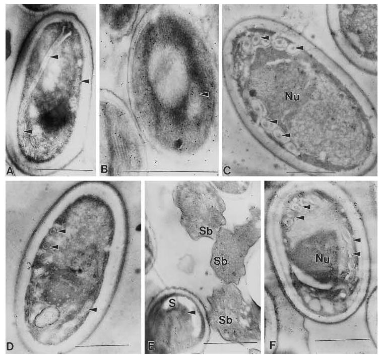
FIG. 2.

Nosema. locustae monoclonal antibody localization with 10 nm gold (Nanoprobes, Inc., Yaphank NY). Monoclonal antibody 19F.9.24 demonstrated a generalized localization to the spores of A, B, and C and reacted with the polar filament (arrow heads) of A and B. Monoclonal antibody 3B1.23 demonstrated a generalized localization to the spores of D, E. and F and reacted with the polar filament (arrow heads) of D and F. Additionally, a generalized localization in the sporoblasts (Sb) of E is noted. A and D are Nosema locustae, B and E are Encephalitozoon cuniculi, and C and F are Pleistophora sp. from the muscle of Callinectes sapidus. Sections through pelleted spores (A. D) and/or spores in host cells (B, C, E and F) contain spores and sporoblasts that are not necessarily visible in the plane of section, but which can bind antibody resulting in gold labeling with the secondary antibody. In uninfected host cells or tissue no antibody binding, gold labeling, is seen (data not shown).
