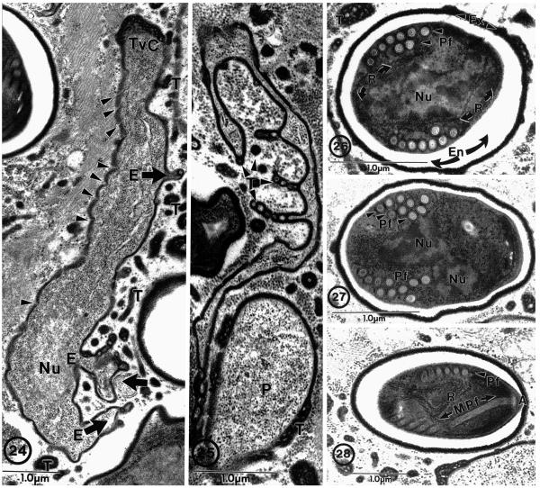
Fig. 24–28.
Brachiola vesicularum from human skeletal muscle biopsy. 24. Proliferative cell with three protoplasmic extensions (E) projecting from the cell (broad arrows). Vesiculotubular appendages (T) associated with elongated cell and protoplasmic extensions. Note the nucleus (Nu), dense plasmalemma (arrowheads), and vesiculotubular cap (TvC) on the end of the parasite cell. 25. Example of elaborate nature of protoplasmic extensions with associated vesiculotubular appendages (T) next to a proliferative (P) cell. 26. Section through a spore illustrating the most common polar filament arrangement observed: nine cross-sections of an anisofilar polar filament (PF) coil arranged in two rows with two to three narrower diameter cross-sections. Spore structures include dense accumulations of ribosomes (R), nuclei (Nu), lucent endospore (En), and dense exospore (Ex) with some appendage material still attached. 27. Spore containing ten polar filament (Pf) cross-sections clustered into three rows (arrowheads). 28. Section through anterior anchoring disc complex (A) of the polar filament (Pf) illustrating its anterior manubroid (Mpf) portion and the cross-sections arranged in a single row. Note the presence of numerous ribosomes (R) inside the spore and the clusters of vesiculotubular material in the host cell cytoplasm. Bar = 1 μm. (All figures on this plate taken from Cali et al. (1998) with permission).

