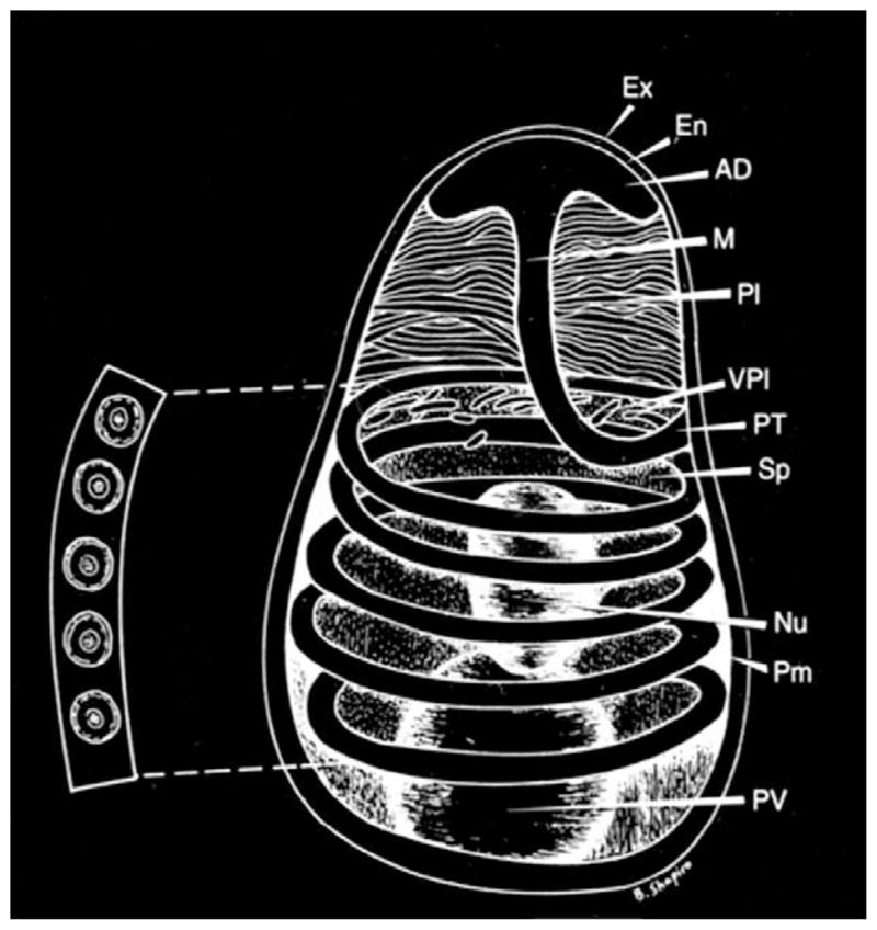
Fig. 1.

Diagram of a microsporidian spore. Spores range in size from 1 to 10 μm. The spore coat consists of an electron dense exospore (Ex), an electron lucent endospore (En) and plasma membrane (Pm). It is thinner at the anterior end of the spore. The sporoplasm (Sp) contains a single nucleus (Nu), the posterior vacuole (PV) and ribosomes. The polar filament is attached to the anterior end of the spore by an anchoring disc (AD), and is divided into two regions: the manubrium or straight portion (M), and the posterior region forming five coils (PT) around the sporoplasm. The manubrium is surrounded by the lamellar polaroplast (Pl) and vesicular polaroplast (VPl). The insert depicts a cross-section of the polar tube coils (five coils in this spore), demonstrating the various concentric layers of different electron density and electron dense core present in such cross-sections. [Reprinted with permission from Wittner, M., Weiss, L.M. (1999). The Microsporidia and Microsporidiosis. ASM Press, Washington, DC].
