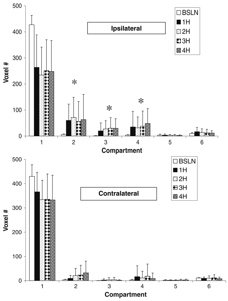
Fig. 3.
Tissue compartmental volumes (voxel #, voxel = 2.57 µl) as illustrated in Fig. 2a for the ipsilateral (top) and contralateral (bottom) hemispheres up to 4H post pMCAO (n=9). Significant (P<0.05) differences were observed between baseline and 1- through 4-h volumes in compartments 2 through 4 in the left hemisphere. In the right hemisphere, significant differences between baseline and hours one through 4 were observed in compartments 2, 3, and 4. *P<0.005 compared to baseline

