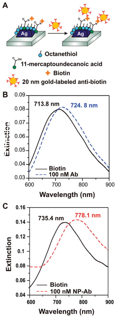
Figure 2.

Experiment schematic and LSPR spectra. (A) Biotin is covalently linked to the nanoparticle surface using EDC coupling agent, and antibiotin labeled gold nanoparticles are subsequently exposed to the surface. LSPR spectra are collected before and after each step. (B) LSPR spectra before (solid black) and after (dashed blue) binding of native antibiotin, showing a Δλmax of 11 nm. (C) LSPR spectra before (solid black) and after (dashed red) binding of antibiotin labeled nanoparticles, showing a Δλmax of 42.7 nm.
