
Figure 2.
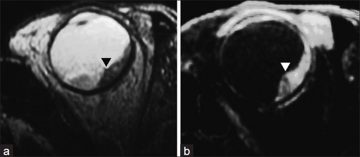
Magnetic resonance imaging shows a plaque-like lesion of uneven thickness along the posterolateral surface of the globe in close relation to the choroid (►) with associated retinal detachment. The lesion is hypointense on T2W study and hyperintense on STIR images