
Figure 4.
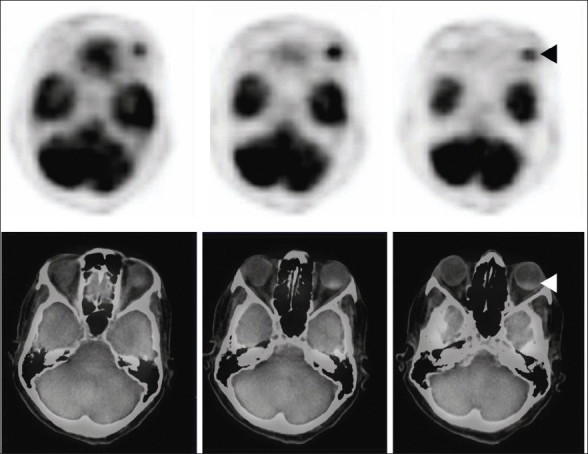
F18 FDG PET CT: Transaxial section across the orbit shows intense focus of FDG activity in the left eye ball posteriorly—fusion image shows coregistration in the posterior choroid corresponding to the radiologic lesion seen in computerized tomogram (◄)