
Figure 1.
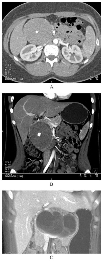
Cross-sectional imaging of a solid-pseudopapillary neoplasm (SPN). (A) Axial CT image of an SPN. (B) The same lesion but from a coronal view. Note that in the arterial phase, this particular lesion is mainly solid. A characteristic central calcification is clearly visible in both views. (C) A coronal CT image of an SPN that is mainly cystic. Note again the calcifications (white arrow).