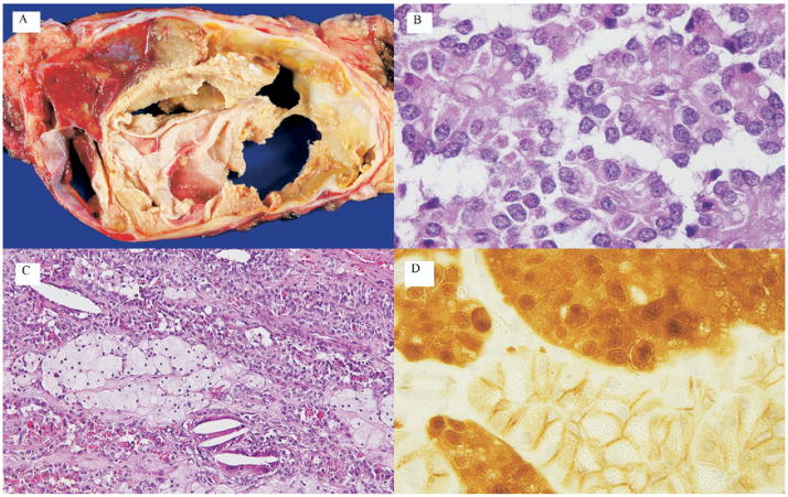
Figure 2.
Gross and histopathologic appearance of solid-pseudopapillary neoplasm (SPN). (A) Gross appearance of an SPN. Note the well-delineated mass with spaces (cysts) containing friable pale tan to yellow material. Hemorrhagic areas can be seen (arrow). (B) On microscopic examination, pseudopapillae are formed by loosely cohesive, monotonous short columnar cells surrounding delicate blood vessels. (C) Histologic patterns are variable. This particular example of an SPN exhibits islands of foamy macrophages (arrow), scattered cholesterol clefts with intracytoplasmic periodic-acid Schiff-diastase resistant hyaline globules. (D) Immunolabeling of the neoplasm for β-catenin exhibits both nuclear and cytoplasmic staining pattern (white arrow). Adjacent normal pancreas exhibits membranous pattern only (black arrow).

