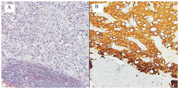
Figure 3.
Solid-pseudopapillary neoplasm metastatic to a lymph node. (A) This lymph node contains metastatic solid-pseudopapillary tumor, leaving only a small contingent of lymphocytes (hematoxylin and eosin). (B) Resident lymphocytes stain pale blue with the counterstain (lower part of field); neoplastic cells exhibit nuclear and cytoplasmic labeling with an antibody for β-catenin.

