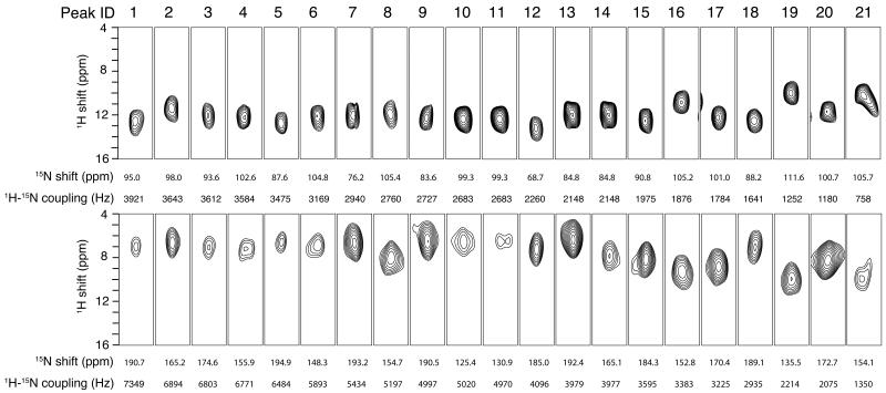
Figure 5.
Strip plots extracted from the three-dimensional HETCOR/SLF spectra (Figure 4A and 4B) obtained for uniformly 15N-labeled Pf1 coat protein in two differently aligned bicelles. Top: The bilayer normals are perpendicular to the magnetic field. Bottom: The bilayer normals are parallel to the magnetic field.

