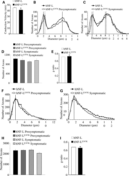
Figure 6.
Reduction of MNCV and reduced axonal diameters in hNF-LE397K mice. (A) MNCV was measured from axons of the sciatic nerve in hNF-L and hNF-LE397K mice. There was a statistically significant difference between conduction velocities from hNF-L and hNF-LE397K mice. Statistical analysis was performed by unpaired t-test. *P< 0.03. (B and C) Distributions of axonal diameters from all axons of the fifth lumbar motor root prior to (B) and after (C) the onset of overt hind limb weakness in age-matched hNF-L and hNF-LE397K mice. The peak diameter for small motor axons was reduced by 0.5 μm prior to the onset of overt hind limb weakness in hNF-LE397K mice (B). After onset, the peak diameter was reduced by 0.5 μm for both small and large motor axons in hNF-LE397K mice (C). Motor axon diameter distributions were analyzed for overall statistical differences utilizing the Mann–Whitney U-test. There was a significant difference in diameter distributions between hNF-L and hNF-LE397K mice for both presymptomatic (P = 0.011) and symptomatic (P< 0.001) time points. (D) The total number of axons in the fifth lumbar motor root for age-matched hNF-L and hNF-LE397K mice. Total axon numbers were analyzed for statistical significance by two-way ANOVA. (E) The ratio of axon diameter/fiber diameter (g-ratio) was analyzed from 10% of all motor axons of symptomatic mice. Axons were randomly selected and localized throughout the entire motor root. g-Ratios were analyzed for statistical significance by the Mann–Whitney U-test. hNF-LE397K g-ratio was significantly larger. (F and G) Distributions of axonal diameters from all axons of the fifth lumbar sensory root prior to (F) and after (G) the onset of overt hind limb weakness in age-matched hNF-L and hNF-LE397K mice. The peak diameter was unaltered prior to or after the onset of hind limb weakness in sensory axons of hNF-LE397K mice. Sensory axon diameter distributions were analyzed for overall statistical differences utilizing the Mann–Whitney U-test. There was a significant difference in diameter distributions between hNF-L and hNF-LE397K mice for both presymptomatic (P < 0.001) and symptomatic (P< 0.001) time points. (H) The total number of axons is reduced in symptomatic hNF-LE397K mice relative to age-matched hNF-L mice. However, the difference did not reach statistical significance. Total axon numbers were analyzed for statistical significance by two-way ANOVA. (I) g-Ratios were analyzed from 10% of all sensory axons of symptomatic mice. Axons were randomly selected and localized throughout the entire motor root. g-Ratios were analyzed for statistical significance by the Mann–Whitney U-test. hNF-LE397K g-ratio was significantly smaller. MNCVs were measured in a minimum of 15 mice per genotype. Each point represents the averaged distribution of axon diameters from the entire roots of at least three mice for each group. g-Ratios were measured on at least three mice per genotype. SEM is reported for all g-ratios. However, they are too small to be seen in the figure. SEM motor: hNF-L = 0.005; hNF-LE397K = 0.004; SEM sensory: hNF-L and hNF-LE397K = 0.002.

