Figure 8.
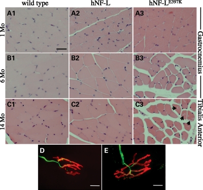
hNF-LE397K mice display progressive muscle atrophy in hind limb muscles without denervation. To determine the morphology of lower hind limb muscles, cross-sections of the gastrocnemius and TA muscles were analyzed from wild-type, hNF-L and hNF-LE397K mice. Muscles were isolated, sectioned and stained with hematoxylin and eosin. (A) Gastrocnemius muscle fibers of 1-month-old hNF-L and hNF-LE397K mice were indistinguishable from wild-type mice. Muscle was analyzed at 1 month to determine if muscle pathology was observed prior to the onset of overt phenotypes. (B) Gastrocnemius fibers of 6-month-old hNF-LE397K mice were atrophied after the onset of hind limb weakness, whereas hNF-L muscle fibers appeared similar to wild-type mice. (C) TA fibers of 14-month-old hNF-LE397K mice were atrophied relative to wild-type and hNF-L mice, suggesting that muscle alterations in the hind limb are not limited to the gastrocnemius. Additionally, the formation of clear inclusions was increased in the muscle of aged hNF-LE397K mice (arrow). Scale bar, 50 μm. (D and E) Gastrocnemius muscle sections were immunostained with antibodies recognizing NFs (α-NF-L) to identify the axon, and fluorescently conjugated α-bungarotoxin to identify the NMJ. There were no differences in the innervation of NMJs in hNF-L (D) and symptomatic hNF-LE397K (E) mice. Scale bar, 10 μm. Muscle and NMJ analysis was performed on four mice per genotype.

