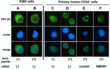
Figure 4.
NmU activates PKc-βII in hematopoietic cells. K562 cells were (A) unstimulated or (B) stimulated with NmU peptide for 30 minutes, and the subcellular localization of PKC-βII was detected by immunofluorescence microscopy. A representative image from 5 different fields of view for each condition is shown. Subcellular localization of PKC-βII was also determined in non-nucleofected primary CD34+ cells that were (C) unstimulated or (D) stimulated with NmU peptide for 30 minutes as well as in primary CD34+ cells that were nucleofected with (E) control or (F) NMUR1 siRNA before NmU peptide stimulation for 30 minutes. As with K562 cells, representative images are shown. PKC-βII in unstimulated control and NMUR1 siRNA-treated cells resided in the cytoplasm as shown in panel C (data not shown).

