FIGURE 8.
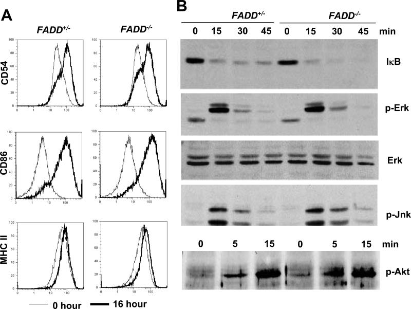
Analysis of activation marker upregulation and intracellular signaling in B cells. (A) Induction of CD54, CD86 and MHC class II was analyzed by staining with indicated antibodies at 0 (thin lines) and 16 h (thick lines) after LPS stimulation of sorted FADD-/- mutant and FADD+/- control B cells. (B) Total proteins from FADD+/- and FADD-/- B cells stimulated with LPS (10 μg/ml) for the indicated times were analyzed for NF-κB activation by detecting degradation of IκB. To analyze Erk activation, the same nitrocellulose membrane was re-probed with antibodies specific for phosphorylated (p)-Erk1/2, and probed with anti-Erk1/2 antibodies after stripping. The membrane was probed for the fourth time with anti-p-Jnk antibodies after the second stripping. A separate membrane with samples collected at the indicated times was probed with anti-p-Akt antibodies to detect Akt phopshorylation induced by LPS stimulation (10 μg/ml). FADD+/- B cells were used as control. Data shown are representative of at least three independent experiments.

