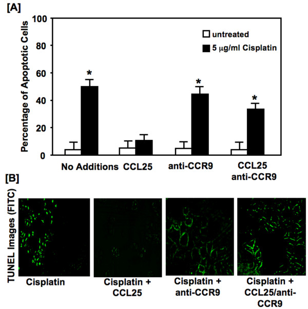
Figure 2.
Cisplatin-induced apoptosis. Panel A: MDA-MB-231 cells were cultured for 24 hours with 5.0 μg/ml of cisplatin with or without CCL25 (100 ng/mL) plus 1 μg/mL of anti-human CCR9 or isotype controls. Cells were stained with annexin V and propidium iodide (PI). Analysis by flow cytometry of the stained cells distinguished apoptotic (annexin V positive) cells from viable (no fluorescence) and necrotic (PI positive) cells. Asterisks (*) indicate significant differences (p < 0.01) between CCL25-treated and untreated BrCa cells. Panel B: MDA-MB-231 cells were cultured for 24 hours with 5.0 μg/mL cisplatin or with 0 or 100 ng/ml of CCL25 plus anti-human CCR9 or isotype control Abs (1 μg/mL). Detection of apoptotic cells was carried out using the terminal deoxynucleotidyl transferase-mediated dUTP nick-end labeling (TUNEL) method. Apoptotic cells exhibited nuclear green fluorescence with a standard fluorescence filter set (520 ± 20 nm).

