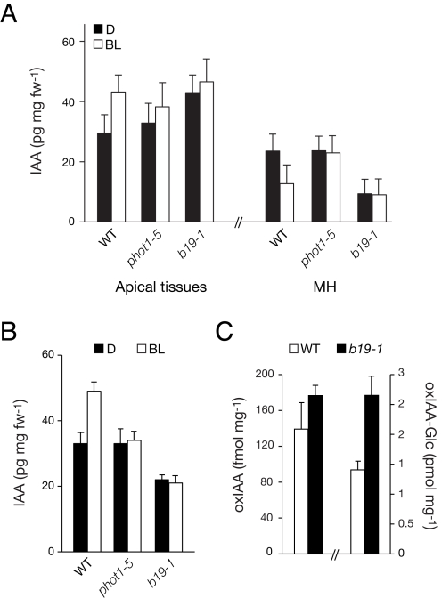
Figure 3. IAA accumulation in dark-acclimated seedlings.
(A) Free IAA levels in wild-type (WT), phot1–5, and b19–1 seedlings. IAA measurements were determined as described [43] after 2.5 h directional blue light (BL, 1 µmol m−2 s−1) or continued darkness (D). Apical tissues including the upper hypocotyl, petioles, and cotyledons were excised for analysis, in addition to the mid hypocotyl (MH). (B) Free IAA determinations as in (A), but after cotyledon excision. Apical tissues including the upper hypocotyl and cotyledonary node with the lower half of the petioles were excised for analysis. (C) Accumulation of auxin catabolites oxIAA and oxIAA-Glc in dark-acclimated wild-type and b19–1 seedlings. Upper hypocotyls/cotyledonary nodes were excised and assayed. In each case, results represent the mean + SD, n = 100 seedlings in three independent experiments.

