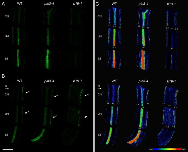
Figure 6. Blue-light-driven lateral auxin gradients in dark-acclimated hypocotyls.
(A) Auxin distributions in dark-acclimated wild-type (WT), pin3–4, and b19–1 seedlings were monitored using the auxin reporter DR5rev:GFP. DR5rev:GFP signals in the cotyledonary node (CN), upper hypocotyl (UH), and elongation zone (EZ). Note the lack of signal in the central vasculature in b19–1. (B) DR5rev:GFP signals in hypocotyls exposed to a directional blue light (BL, 1 µmol m−2 s−1) for 3 h. In all genotypes, signals are asymmetrically localized in the epidermal cells on the shaded side of the hypocotyl (white arrows) in both the CN and UH. (C) Heat map representation of DR5rev:GFP signals shown in (A) and (B). Quantification of the mean pixel intensity of the DR5rev:GFP signals associated with the epidermal regions of the CN are shown, as are the central vasculature signals for the CN and UH. Quantification was performed using the analyze and measure functions within ImageJ (version 1.43u). Data are representative of n = 20 seedlings in three independent experiments for wild type and n = 10 in two separate experiments for pin3–4 and b19–1. Scale bar = 200 µm.

