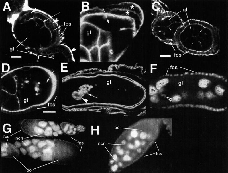
Figure 5.
Genes required for mid-oogenesis. (A) fs(1)186/fs(1)186 mutant's egg chamber labeled for actin to show abnormal aggregation of follicle cells over the oocyte. The arrowheads point out some of the follicle cells that do not contact the germline. (B) Stage 9 egg chamber from fs(1)186/fs(1)186 in which follicle cells have formed a two-layer epithelium over the oocyte (* indicates follicle cell layers). (C) The follicle cell aggregation phenotype is partially suppressed in fs(1)186/fs(1)186; Bic-DPA66/Df(2L)TW119. (D,E) Actin staining in a stage 5 (D) and a stage 9 (E) fs(1)234 /fs(1)234 mutant egg chamber showing the progressive loss of germline cell membranes. (F) Same egg chamber as in (E) stained for nuclei. The strong actin staining in (E) is due to aggregation of ring canals (arrow in E) and the border cells (arrowhead in E and F). Scale bars in (A,C,D) = 20μm. Nuclear labeling of fs(1)225/fs(1)225 mutant ovaries reveals (G) enlarged nurse cell nuclei and (H) supernumerary nurse cells.

