
Figure 6.
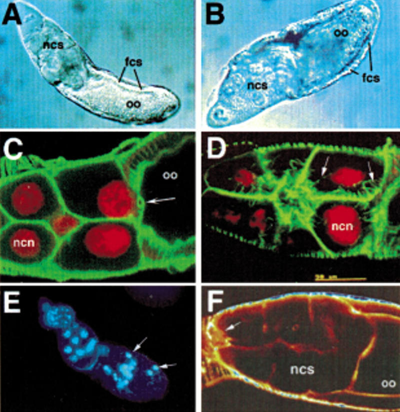
(A -D) Defects in late transport of nurse cell contents in mutants for fs(1)140 and fs(1)3. oo, oocyte; fcs, follicle cells; ncs, nurse cells; ncn, nurse cell nucleus. (A,B) Nomarski views of (A) fs(1)140 and (B) fs(1)3 egg chambers in which nurse cell dumping has failed. (C,D) Double labeling of nuclei (red) and actin (green) in (C) fs(1)140/fs(1)140 and (D) fs(1)3/fs(1)3. In fs(1)140/fs(1)140, actin cables fail to form and the nurse cell nuclei appear to become caught in the ring canals (arrow) during dumping. In fs(1)3/fs(1)3, actin cables form normally (arrows point to actin cables). (E) Nuclear staining of fs(1)164/fs(1)164 reveals pycnotic nuclei (arrows) in stage 8 of oogenesis. (F) Actin staining of fs(1)221a/fs(1)221a reveals failed border cell migration (arrow points to border cells).