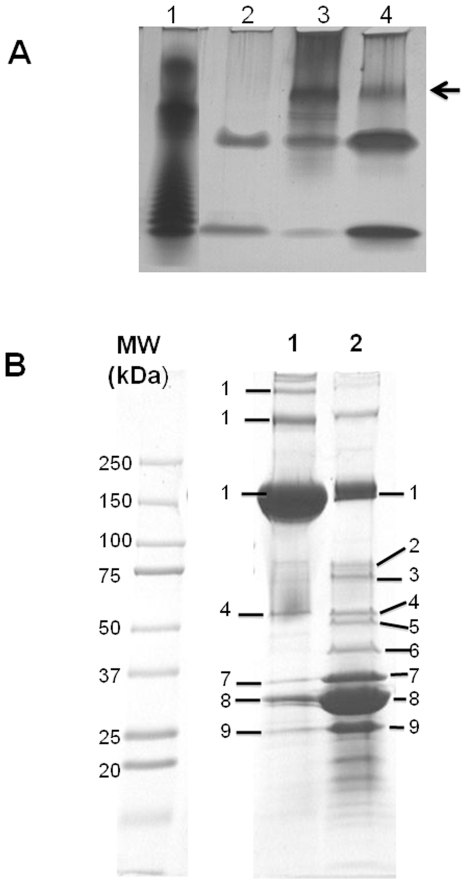
Figure 3. Analysis of lipopolysaccharide and proteins in nanopods and outer membrane.
Panel A. Analysis of lipopolysaccaride (LPS) by tricine-SDS-PAGE and silver staining. Samples of LPS were extracted from: purified nanopods (Lane 2), Delftia sp. Cs1-4 cells grown on phenanthrene (Lane 3) and Delftia sp. Cs1-4 cells grown on pyruvate (Lane 4). Lane 1 is an LPS standard (Salmonella typhimurium). The arrow indicates a dense band of LPS present in whole cell preparations but absent from nanopods. All samples were loaded at the same dry weight (200 µg). Panel B. SDS-PAGE Protein profiles of nanopods (Lane 1) and the outer membrane (OM) of phenanthrene-grown Delftia sp. Cs1-4 cells (Lane 2). Proteins identified in gel slices from the OM sample are (locus in Delftia sp. Cs1-4): 1. NpdA (DelCs14_2799), 2. Fiu-like TonB-dependent siderophore receptor (DelCs14_0908), 3. TonB-dependent siderophore receptor (DelCs14_5618) 4. Protein with domain of unknown function 1302 (DelCs14_4425), 5. RND efflux system, outer membrane lipoprotein (DelCs14_5845) 6. 4. Type II L-Asparaginase, 7. OmpC-like protein (DelCs14_0125), 8. Omp32 (DelCs14_0124). 9. OmpA-like protein (DelCs14_3211).

