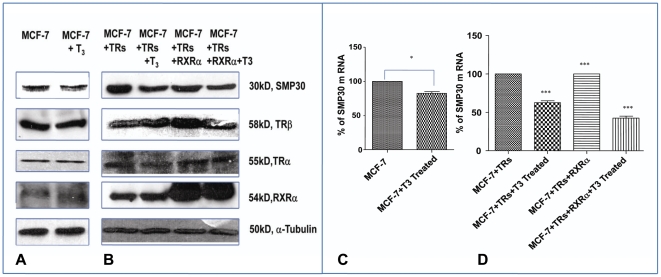
Figure 1. Downregulation of SMP30 gene in MCF-7 cell line by 3, 3′5 triiodo L thyronine.
(A) Representative Western blot of total SMP30 protein in response to overnight treatment with T3 in MCF-7 cells. (B) Expressional analysis of SMP30 gene after overnight T3 treatment to TRs or TRs and RXRα cotransfected MCF-7 cells in Fig.1B was performed by Western blot analysis. 100 µg of protein from whole cell extract was used for Western analysis.In all figures (1A and B) SMP30 protein was detected by SMP30 antibody at ∼30 kDa, TRβ protein was detected by TRβ antibody at 58 kD, TRα protein was detected by TRα antibody at 55 kD and RXRα protein was detected by RXRα antibody at 54 kD were assessed. α-Tubulin was used as a loading control. In Fig. 1C and D, total RNA from cells were harvested and analyzed by quantitative RT-PCR. Shown are the mean values from triplicate samples normalized to GAPDH. CT values obtained from the real time PCR was used to compare the expression label of treated sample from control assuming 100% amplification. Paired student's t test performed; *, P<0.01 results were confirmed in three independent experiments as in Fig. 1C. *** P<0.0001difference from control using ANOVA as in Fig. 1D.

