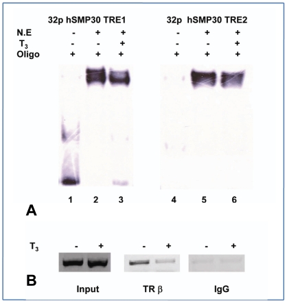
Figure 3. Effects of T3 on TR-DNA interaction.
(A) Electrophoretic mobility shift assay for hSMP30 TRE 1 and 2 in presence of T3 hormone. Lane 1, 4 are labeled hSMP30 TRE1 and 2 respectively. Lane 2, 5 are labeled TREs with 10 µg of N.E., Lane 3, 6 are labeled TREs with 10 µg of N.E in presence of 1 µM T3 hormone. (B) TRβ binding to SMP30 promoter is affected by T3 treatment was shown in ChIP. After 1 hr. treatment with 1 µM T3, MCF-7 cells were crosslinked with 1% formaldehyde and ChIP assays were performed according to manufacturer's protocol (Upstate Biotechnology) with some minor modifications. After reverse crosslinking by heating the samples at 65°C for 4–6 hrs and treating with proteinase K, DNA was elute by phenol chloroform extraction then ethanol precipitation. PCR was performed to visualize the enriched DNA fragments. In vivo association of TRβ protein complex with the hSMP30 promoter was demonstrated by the amplification of hSMP30 TREs specific DNA fragments from chromatin complexes precipitated by antibodies for TRβ.

