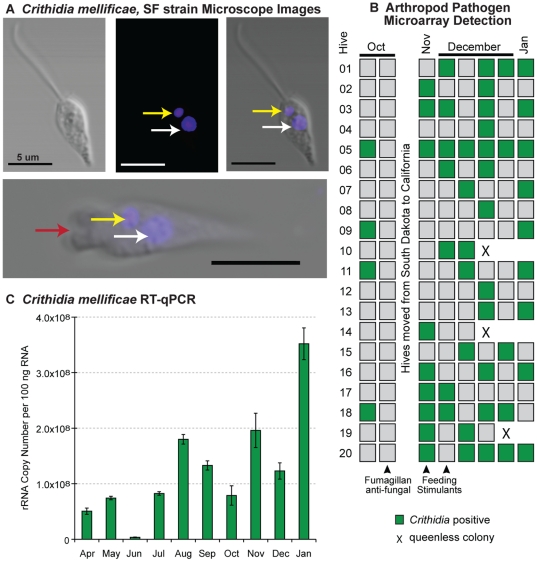
Figure 6. Crithidia mellificae, SF strain detection and quantification.
(A) Light and fluorescent microscope images illustrate key features of this trypanosomatid parasite including DAPI stained kinetoplast DNA (yellow arrow) and nuclear DNA (white arrow), as well as the flagellar pocket (bottom panel, red arrow); scale bar = 5 µm. (B) Arthropod pathogen microarray detection of Crithidia mellificae in each colony (5 bees per sample) from October 2009 to January 2010. (C) Relative abundance of Crithidia mellificae throughout the time-course as assessed by RT-qPCR of pooled monthly time-course samples; quantification of rRNA copy number based on a standard curve as described in Materials and Methods.

