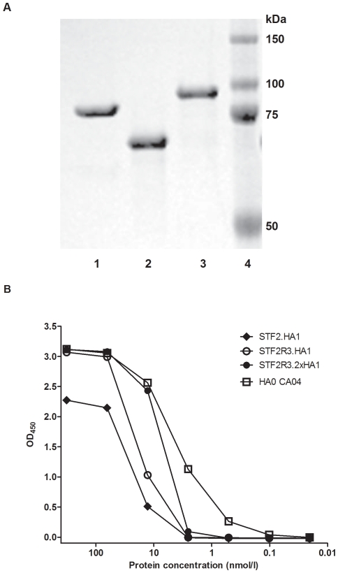
Figure 2. SDS-PAGE and antigenicity analyses of three purified recombinant. vaccine candidates.
(A) Purified recombinant proteins were separated on an 4–12% SDS PAGE (0.5 µg protein/lane) and stained with Coomassie Blue. Lane1: STF2.HA1; lane 2: STF2R3.HA1; lane 3, STF2R3.2x HA1; and lane 4, Protein Marker. (B) Reactivity of ferret post infection serum to various vaccine candidates or reference antigen. ELISA plates were coated with serially diluted various CA07 proteins or HA CA04 (Protein Sciences) starting at 320 nmol/l in in PBS, reacted to ferret anti-CA7 serum, and detected with HRP-conjugated goat anti-ferret IgG. Mean OD450 of triplicates was read and graphed.

