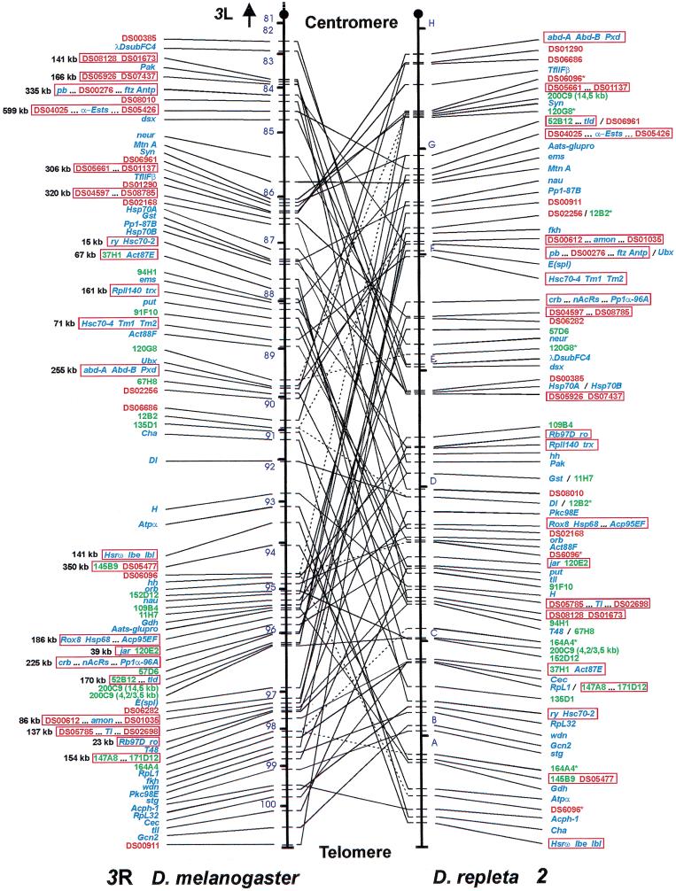
Figure 1.
Large-scale comparison of the gene organization of Muller's element E between Drosophila melanogaster and Drosophila repleta. Connecting lines match the cytological position of orthologous markers. Dashed lines point out those markers whose location can be assigned to a single chromosomal site in D. melanogaster but that produce more than one hybridization signal in D. repleta. Mapped markers are indicated in blue (genes), green (cosmids), or red (P1 phages). When two or more markers provide redundant mapping information, only representative markers are shown. For precise mapping location of all the markers in D. melanogaster and D. repleta, see supplemental Table 1 (available on-line at http://www.genome.org). Red open rectangles indicate the 21 conserved segments comprising at least two independent (nonoverlapping) markers that presumably represent ancient physical associations not disrupted during the evolution of the two compared lineages. Their estimated sizes in D. melanogaster are indicated. Two genes localized by other authors, Hsrθ (Peters et al. 1984) and orb (H. Naveira, pers. comm.), are also included. Cosmid 200C9 does not appear as a single marker but as two sets of independent subclones (Ranz et al. 1999). Chromosomal arm 3R of D. melanogaster is partitioned into 20 (from 81–100) out of 102 numbered sections in which the D. melanogaster genome is subdivided (Bridges 1935). Chromosome 2 of D. repleta shows its seven main lettered sections according to Wharton (1942).

