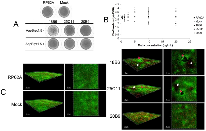
Figure 3. Anti-AapBrpt1.5 MAbs affected biofilm formation by S. epidermidis.
(A) Macroscopic profiles of the biofilms cultured in polystyrene plates. The biofilms were cultured in TSB medium containing each MAb (10 µg/mL, 0.07 µM) alone or both MAbs (10 µg/mL, 0.07 µM) and AapBrpt1.5 (untagged, 3.2 µg/mL, 0.14 µM). The images represent one of three independent experiments. (B) Anti-AapBrpt1.5 MAbs affected biofilm formation by S. epidermidis RP62A in a dose-dependent manner. The biofilm formation was measured using crystal violet staining, and the results are depicted as means ± SD of three independent experiments. (C) Three-dimensional structures of 14-h-old biofilms. Biofilm formation by S. epidermidis RP62A in the presence of each MAb (10 µg/mL) was visualized using Live/Dead viability staining (SYTO9/PI) and observed under a confocal laser scanning microscopy (CLSM). Green fluorescent cells are viable, whereas red fluorescent cells are dead. The images, representing one of three independent experiments, were three-dimensionally reconstructed using Imaris software (Bitplane, http://www.bitplane.com) based on CLSM data at approximately 0.5 µm increments. “RP62A”: untreated, “Mock”: normal mouse IgG-treated; white arrows and red arrows indicate thin areas and crater-like micropores, respectively.

