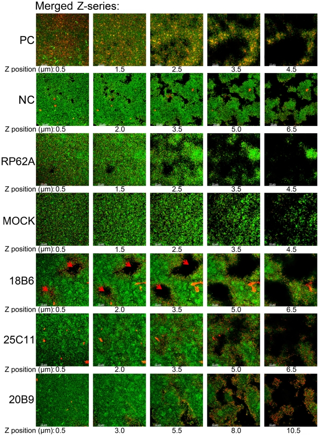
Figure 7. Aap expression in biofilms of S. epidermidis.
Aap in the biofilms of S. epidermidis RP62A was probed with MAb25C11 (10 ng/mL) and Cy3-conjugated secondary antibody (1∶100 diluted, red fluorescence), and the bacteria were further stained with SYTO9 (1 µM, green fluorescence). Aap expression was observed under a Leica TCS SP5 CLSM. Confocal microscopy Z-series of the biofilms were acquired in 0.5-µm increments. “PC”: positive control (antigens contained in the biofilm were probed using mouse anti-S. epidermidis serum (1∶400 diluted) and Cy3-conjugated secondary antibody, showing that antibodies could diffuse to the inner side of the biofilm), “NC”: negative control (the biofilm formed in the presence of MAb25C11 was probed with Cy3-conjugated secondary antibody alone to establish that the MAb-treated biofilms no longer contained initially added MAb (10 µg/mL), that could cause false-positive immunofluorescence, after 14 h culture), “RP62A”: untreated, “Mock”: normal mouse IgG-treated, the red arrow indicates the crater-like micropores.

