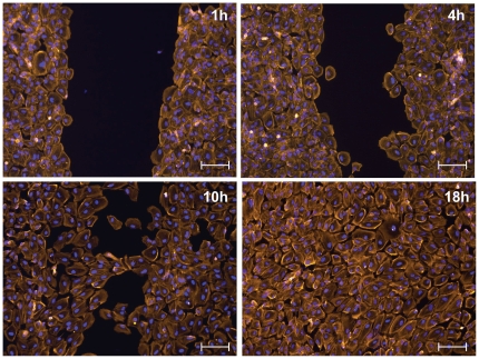
Figure 4. Fluorescence microscopy images of epithelial cells after removal of Parafilm™ inserts: Epithelial cells (ARPE-19) were cultured in stripe patterns.
The Parafilm™ stripes were removed enabling cells to re-populate the free surface created by removal of the parafilm. Cells were fixed at 1,4, 10 and 18 hours after insert removal and stained with DAPI (blue) to show the nucleus and with phalloidin (orange) to show the F-actin cytoskeleton. The scale bar for each image is 100-µm wide.

