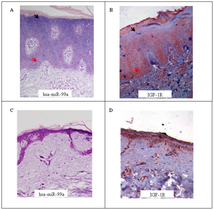
Figure 4. Reciprocal expression of IGF-1R and hsa-miR-99a.
The distribution of hsa-miR-99a in psoriatic lesional skin (A) or normal skin (C) was assessed by in-situ hybridization. Paraffin-embedded sections of lesional skin biopsies were hybridized with an LNA probe specific to hsa-miR-99a. The cytoplasm of positive cells was stained blue (indicated by black arrows). Nuclei were stained with Nuclear Fast Red counter stain (indicated by red arrows). Magnification, 200x. (B) and (D) The same paraffin-embedded skin biopsy shown in (A and Crespectively;) was immunostained with anti-IGF-1R antibodies. Antibody binding was visualized with the substrate-chromogen AEC. IGF-1R positive cells were stained red (indicted by the red arrow). Nuclei were stained blue with hematoxylin (indicated by the black arrow). Magnification, 200x.

