
Fig. 5.
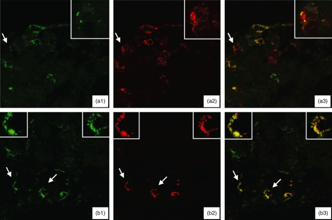
Immunofluorescence laser scanning confocal microscopy studies of skin specimens from two patients with bullous pemphigoid (a, b) showing staining for the specific eosinophil marker CD 125 (green; a1 and b1), tissue factor (red; a2 and b2) and the co-expression of tissue factor and CD 125 (merge; a3 and b3). Particulars of the cells indicated by arrows are shown in the insets. Most of the inflammatory cells co-expressed tissue factor and CD 125 (a3 and b3), thus indicating that they are eosinophils; a few cells expressed either tissue factor or CD 125 alone, thus indicating that the former are inflammatory cells other than eosinophils, and the latter eosinphils that are probably insufficiently activated to express tissue factor.