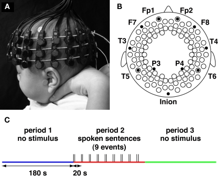
Figure 1.
Experimental settings. (A,B) The NIRS probe and 94 measurement channels. Probes were placed over the bilateral frontal, temporal, temporoparietal, and occipital regions of infants. The probes and measurement channels (open circles in B) were correctly positioned utilizing the 10–20 system. (C) Three periods during the measurement. During the first 3 min, we measured spontaneous brain activation (period 1). After period 1, we provided stimuli by playing Japanese sentences; the sentences were played at 20-s intervals for 3 min (period 2). Finally, we measured brain activation for 3 min without providing the stimulus (period 3), as in period 1.

