Fig. 4.
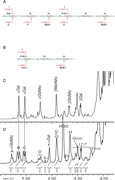
Comparison of the repeating unit structures of the Bacillus SCWP and the anomeric regions of the 600-MHz proton NMR spectra for the SCWP. (A) The structure of the repeating unit of the B. cereus G9241 SCWP; (B) structure of the repeating unit from the B. anthracis SCWPs. The SCWP from all the B. anthracis strains studied to date (Ames, Pasteur and Sterne) have identical structures (Choudhury et al. 2006); (C) 1H-spectrum, anomeric region, for B. anthracis Sterne 7702 SCWP; (D) spectrum for the B. cereus G9241 SCWP. The stoichiometry of the anomeric signals for the B. cereus G9241 strain is indicated. Residue E, anomeric signals attributed to the reducing-end GlcNAc residue. A small signal at approx. δH 5.4 arises from a contaminating α1 → 4 linked glucan.

