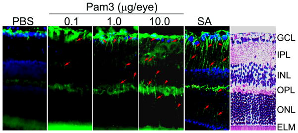
Figure 1. S. aureus and TLR2 agonist enhance GFAP levels in mouse retina.
C57BL/6 mice were given intravitreal injections of PBS (vehicle), TLR2 agonist (Pam3Cys) or S. aureus (SA). After 16 h eyes were enucleated, embedded in OCT and cryosections were stained with rabbit polyclonal anti-glial fibrillary acidic protein (GFAP) antibody. Immunoreactivities are increased in Pam3 or SA challenged retina compared with the PBS controls. Moreover, Muller cell bodies and radial running processes (arrowheads) in the INL and ONL were immunopositive. H & E staining was used as a guide to the retinal layers. GCL, ganglion cell layer; IPL, inner plexiform layer; INL, inner nuclear layer; OPL, outer plexiform layer; ONL, outer nuclear layer; ELM, external limiting membrane.

