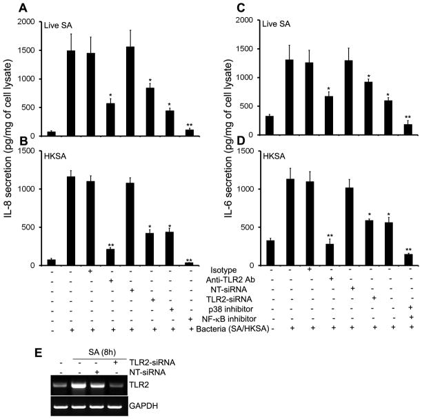
Figure 6. S. aureus induced inflammatory response is attenuated by inhibition of TLR2 and NF-kB signaling.
MIO-M1 cells were pretreated for 1h with isotype (10 μg/ml), anti-TLR2 (10 μg/ml), p38 MAPK (SB, 5 μM) and NF-κB (IS, 25 μM) pathway inhibitors followed by challenge with (A & C) live S.aureus (SA) or (B & D) heat-killed S.aureus (HKSA). After 8h of stimulation culture supernatants were collected and IL-8 & IL-6 levels were quantitated by ELISA. For siRNA transfection, MIO-M1 cells were transfected with non-targeted siRNA (NT-siRNA) or TLR2-siRNA for 48 h, followed by challenge with SA or HKSA for 8h. TLR2 knockdown was assessed by RT-PCR (E). Data are means ± SD of triplicate cultures and are representative of three independent experiments. Statistical analysis was performed using Student’s t-test and indicated statistical differences are comparisons of isotype vs TLR2 antibody, NT-siRNA vs TLR2-siRNA, and untreated vs inhibitor treated cells: *p < 0.05; **p < 0.001.

