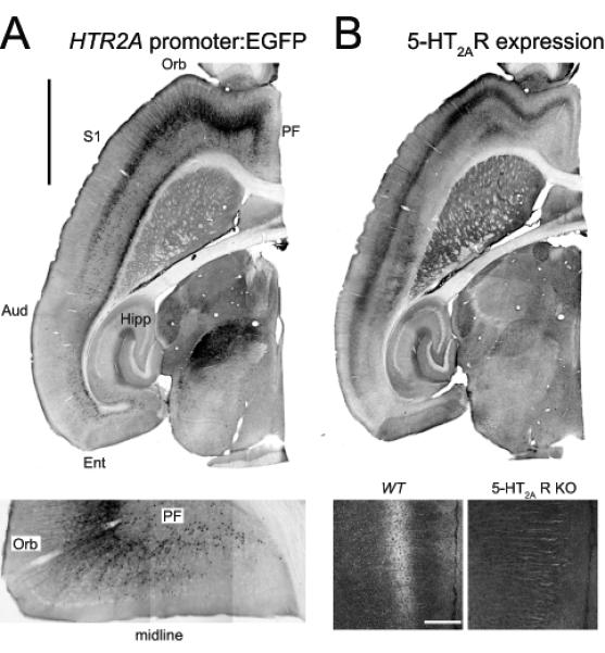
Figure 1. 5-HT2A receptors expression in the cerebral cortex.

A. Expression of the 5-HT2A receptor gene (HTR2A) in the mouse forebrain as revealed in a BAC transgenic mouse expressing EGFP under the control of the 5-HT2A receptor promoter. Calibration bar: 2 mm. Aud: auditory cortex, Ent: entorhinal cortex, Orb: orbital cortex, PF: prefrontal cortex, S1: primary somatosensory cortex. Lower panel. Close up image depicting EGFP expression in an horizontal section through the prefrontal and orbital cortices. B. 5-HT2A receptor expression as determined using an affinity purified anti-5-HT2A receptor antibody. Notice the strong correspondence in EGFP in panel A and 5-HT2A receptor expression in Panel B. Lower panel: 5-HT2A receptor staining in the prefrontal cortex of a wild type (wt) mouse and a 5-HT2A receptor knockout (5-HT2AR KO) mouse. Calibration bar 200 μm. (Modified from Weber and Andrade, 2010).
