Figure 5.
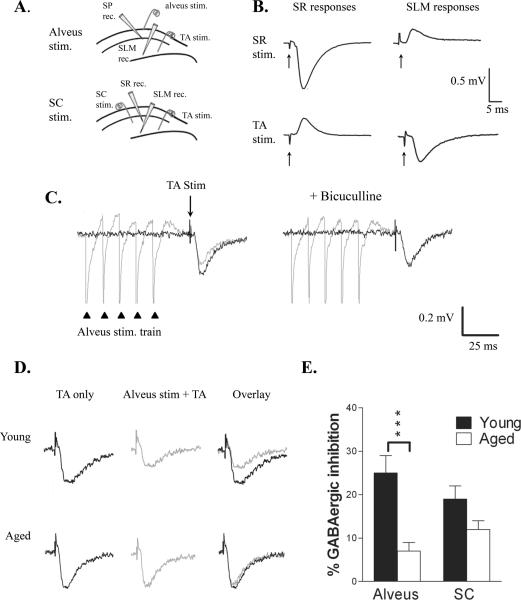
Feedback inhibition of TA input was impaired with aging. A. Diagram of electrode placement used to record TA-evoked fEPSPs in SLM. Inhibition of the TA-evoked response was produced by stimulation in the alveus (top) or SC (bottom). Alvear-evoked responses were additionally recorded in SP, whereas SC-evoked responses were also recorded in SR. B. Field EPSPs recorded in SR and SLM demonstrating isolation of the TA and SC-evoked responses. Arrows indicate stimulus artifacts. C. Left: The TA-evoked fEPSP recorded in SLM (black trace) was inhibited by alvear stimulation (100 Hz, 5 pulses; gray trace) delivered 20 ms earlier. Right: Addition of bicuculline blocked this inhibition. Triangles indicate alvear stimuli; arrow indicates TA stimulus. D. Left: TA stimulation evoked a fEPSP in both young (upper) and aged (lower) rats. Center: Preceding this TA stimulus by 20 ms with alvear stimulation (100 Hz, 5 pulses) inhibited the TA-evoked fEPSP to a greater extent in young than aged rats, as can be seen when the fEPSPs are superimposed (Right). E. Alvear-evoked inhibition was significantly reduced in aged animals (n = 7; P < 0.001), whereas SC-evoked inhibition (n = 7 young, 5 aged) was not different.

