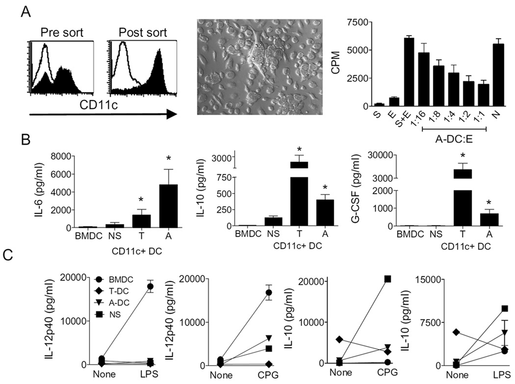
Figure 1. CD11c+ cells derived from the ovarian-cancer microenvironment demonstrate an immune suppressive phenotype.
Panel A shows histograms of Ficoll-purified tumor-associated mononuclear cells (left panel, Pre-sort) and CD11c-sorted tumor-associated DC (right panel, Post-sort). Middle panel shows photomicrograph of CD11c-enriched cells adherent on glass cover slips. Rightmost panel shows tritium incorporation (CPM, counts per minute) into effector (E) cells in response to MHC mismatched stimulators (S) with varying concentrations of ascites-derived dendritic cells (A-DC). N=bone marrow derived DC at 1:1. Panel B shows concentrations of IL-6, IL-10 and G-CSF in the supernatants following short-term incubation of BMDC, splenic DC (NS), tumor-derived DC (T-DC), and ascites-derived DC (A-DC). Each bar is the mean and s.e.m. of six determinations and representative of three separate experiments. Panel C shows concentrations of IL-12p40 (left 2 panels) and IL-10 (right 2 panels) in the supernatants following short-term incubation of 5 × 10⁵ BMDC, NS, T-DC, and A-DC with or without LPS and CpG. Each bar is the mean and s.e.m. of two determinations and representative of three separate experiments.

