
Figure 2.
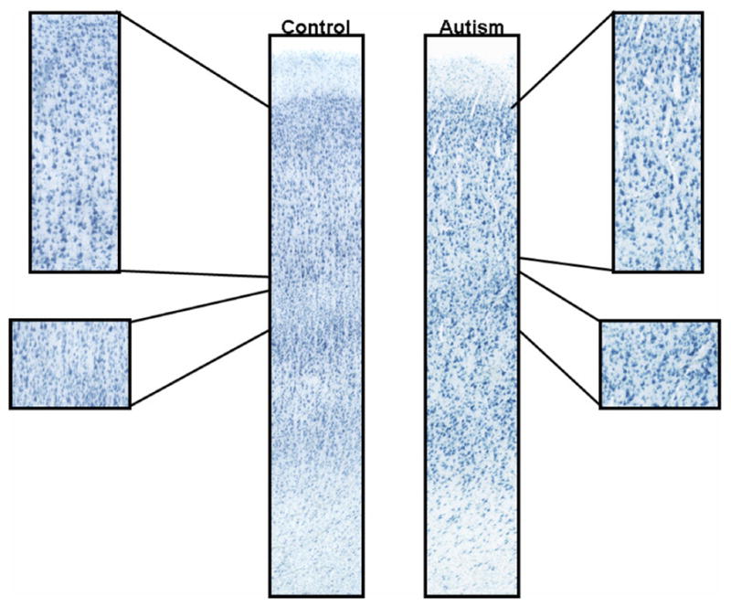
Representative samples of immunohistochemical staining in the PCC and FFG. Control cases from the PCC (a; case 1649; 71.3 months in fixative) and FFG (c; case 4916; 31.9 months in fixative) are on the left and autism cases from the PCC (b; 4999; 13.5 months in fixative) and FFG (d; 5027; 21.7 months in fixative) are on the right. Note that staining is similar in all examples regardless of the fixation time.