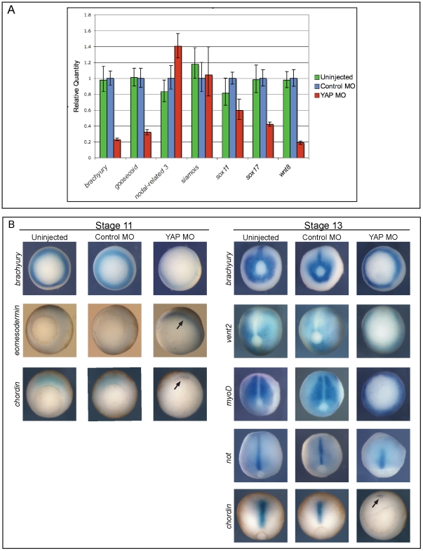
Figure 2. Germ layer markers are expressed in YAP morphant Xenopus embryos, but are temporally delayed.
(A) qPCR analysis of mRNA from uninjected, control MO-injected, and xYAP MO-injected Xenopus embryos collected when controls reached stage 10.5/11. brachyury, goosecoid, wnt8, sox11, and sox17 mRNA levels were reduced, nodal-related 3 (nr3) mRNA levels were increased and siamois mRNA levels remained unchanged in xYAP morphant embryos. (B) In situ characterization of mesoderm gene expression in uninjected, control MO-, and xYAP MO-injected Xenopus embryos. xYAP morphant embryos express each gene in the correct location, but the spatial pattern resembles an earlier developmental stage. For example, brachyury expression in the stage 11 YAP MO embryos is only faintly detected and brachyury expression in the stage 13 YAP MO embryo is indistinguishable from the control stage 11 pattern. chordin expression in the stage 13 YAP MO embryo remains confined to the dorsal blastopore lip (arrow), as is normal at stage 11; it has not elongated with the axial mesoderm as is normal at stage 13. eomesodermin expression in the stage 11 YAP MO embryo remains on the surface in the uninvoluted mesoderm (arrow), whereas in controls, eomesodermin-expressing cells have migrated internally [21]. In the stage 11 panel, all views are vegetal; in the stage 13 panel, the views of brachyury and vent2 embryos and of the YAP MO chordin embryos are vegetal and the remainder are dorsal.

