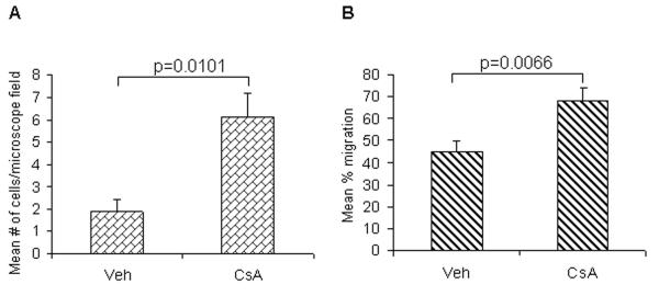
Fig 4.
Effects of CsA treatment on the (A) invasion and (B) wound healing capabilities of A431 cells. Cell invasion was measured with a blind well chemotaxis invasion chamber. 100μl of DMEM containing 20% fetal bovine serum was added into the lower chamber and covered with an 8μm pore size polycarbonate membrane filter. Cells (2.5 × 10⁴) in 100μL of serum free DMEM were seeded into the upper chamber. After incubating for 24 hours at 37°C, membranes were collected and non-invading cells were removed from the upper surface of the membrane using a cotton swab. The membranes were then fixed with methanol, stained with Harris hematolxylin, and photographed microscopically with a 10X objective. The number of invading cells on the lower surface of the membrane was counted in 10 fields per membrane. For wound healing assay, A431 cells were seeded in six-well plates and incubated overnight in starvation medium. Then, the cell monolayers were wounded with a sterile 100-μl pipette tip, washed with starvation medium to remove detached cells from the plates. Cells were left either untreated or treated with CsA, and kept for 24 hrs in CO2 incubator. After 24 hrs, medium was replaced with PBS and cells were photographed using an Olympus (Japan) IX-TVAD digital camera connected to an Olympus IX70 phase-contrast microscope (4X objective).

