
Fig. 1.
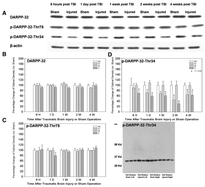
Timecourse of TBI effect on DARPP-32 protein expression and phosphorylation state in rat striatum (N = 6 per group at each timepoint). A, representative western blot timecourse of DARPP-32, p-DARPP-32-Thr75, and p-DARPP-32-Thr34 protein expression. B, optical density of DARPP-32 showing no change in DARPP-32 protein expression at any examined timepoint post TBI. C, optical density of p-DARPP-32-T75 showing no significant change in p-DARPP-32-T75 following TBI. D, optical density of p-DARPP-32-Thr34 showing a significant reduction in p-DARPP-32-T34 in the striatum ipsilateral to injury at all timepoints examined and in the striatum contralateral to injury at 1 day-4 weeks post injury. **Inset image: control western with p-DARPP-32-Thr34 antibody demonstrating a single band on rat striatal tissue. *p ≤ 0.05 normalized to β-actin and compared to sham; ANOVA. Data represented as a percentage of sham following normalization to β-actin ± S.E.M. Abbreviations: SC = sham contralateral to injury; IC = injured contralateral; SI = sham ipisilateral to injury; II = injured ipsilateral; S = sham; I = injured.