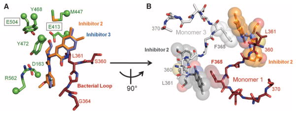
Fig. 2.
Potent β-glucuronidase inhibitors. (A) Crystal structures of Inhibitors 2 and 3 bound to the active site of E. coli β-glucuronidase. (B) Inhibitors are observed to stack cooperatively between monomers in the E. coli β-glucuronidase tetramer. Amino acid abbreviations: D, Asp; E, Glu; F, Phe; G, Gly; L, Leu; M, Met; R, Arg; S, Ser; Y, Tyr.

