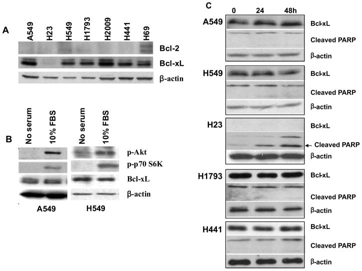
Figure 2.
Effects of PI3K/Akt inhibition on expression of Bcl-xL in lung adenocarcinoma cell lines. (A) Immunoblot analysis of Bcl-2 and Bcl-xL proteins in lung adenocarcinoma cell lines. H69 is a small cell lung cancer cell line and was used as a positive control for Bcl-2 expression. Total cell lysates obtained from the cells maintained in normal 10% serum growth condition were immunoblotted with anti-Bcl-xL, anti-Bcl-2 and anti-β-actin antibodies. (B) Bcl-xL expression is independent of serum concentrations in A549 and H549 cells. Cells were cultured in serum-free media overnight followed by 10% serum for 24h. Immunoblot analysis was used to determine the expression level of Bcl-xL and phosphorylation status of Akt and p70S6K. (C) Bcl-xL expression is independent of PI3k inhibition in lung adenocarcinoma cells. Cells were incubated for 24–48 hours with 25 μM LY294002 and then cell lysate were immunoblotted with antibody against Bcl-xL and PARP.

