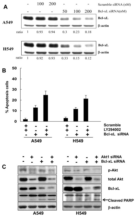
Figure 3.
Apoptotic response induced by the inhibition of PI3K/Akt and Bcl-xL in A549 and H549 cells. (A) Dose-dependent Bcl-xL downregulation in response to Bcl-xL siRNA for 48 hours; expression levels were determined by immunoblot analysis. Results represent one of three experiments with similar results. (B) Bcl-xL gene silencing restores the sensitivity of A549 and H549 cells to PI3K inhibition-induced apoptosis. Percentage of apoptotic cells (sub-G1 population) detected by FACS analysis is represented after cells were transfected with 100 nM Bcl-xL siRNA or scramble siRNA control for 48 hours, followed by 25 μM LY294002 treatment for another 48 hours in the presence of 10% FBS. Knockdown of Bcl-xL alone induces a significant percentage of cells to undergo apoptosis. The combination of Bcl-xL knockdown and LY294002 treatment increased the apoptotic response in a synergistic manner. (C) Effects of combinatorial treatment of Akt1 and Bcl-xL siRNA on phosphorylated Akt (s473), total Akt, Bcl-xL and PARP in A549 and H549 cells. Cells were transfected with double-stranded Akt1 siRNA and/or Bcl-xL siRNA. After an initial transfection (5 h), cells were left untreated for an additional 48 h, and protein lysates were obtained for Western Blotting to measure the expression of Akt phosphorylation at Ser473, total Akt, Bcl-xL and cleaved PARP. (D) A representative histogram of cell cycle distributions and increase of cells in sub-G1 phase (percentage of subG1 fraction is shown in %) induced by combined Bcl-xL and Akt1 siRNA treatment for 48 h in H549 and A549 cells is shown. (E) Apoptosis induced by combined treatment with ABT-737 (8 μM) for 48h or with the enantiomer as negative control (ABT-ctl) and LY294002 (25 μM) in A549 and H549 cells.


