FIGURE 3.
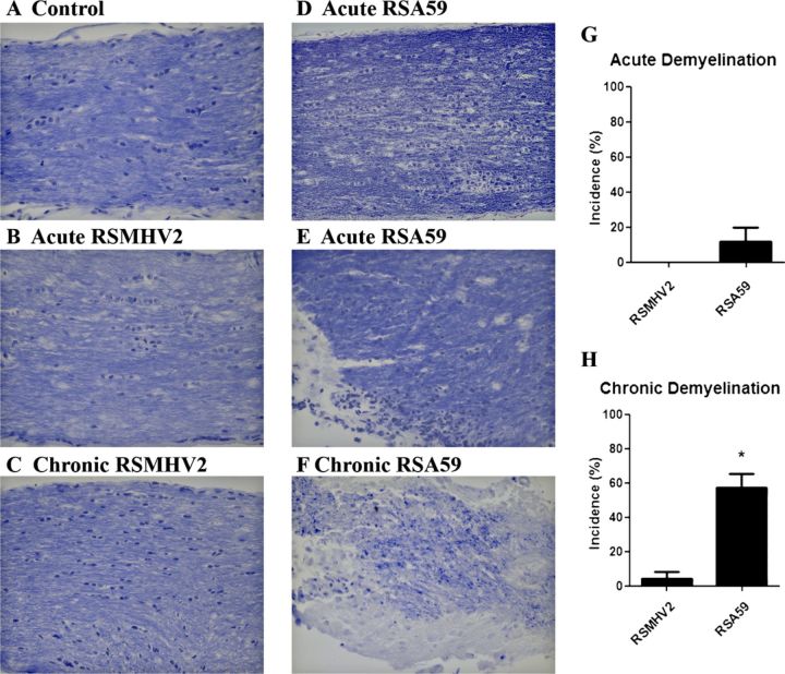
Demyelination in optic nerves of RSA59- and RSMHV-2-infected mice. (A-F) Longitudinal sections of optic nerves from mock- (A), RSA59- (D-F), and RSMHV2 (B, C)-infected mice at days 6 and 30 post inoculation (pi) stained with Luxol fast blue. There is normal myelin staining in the control optic nerve (A). Almost all of the optic nerves in RSMHV2-infected mice had normal myelin at day 6 (B) and at day 30 (C) pi. During acute infection at day 6 pi, the optic nerves in most RSA59-infected mice maintained normal myelin (D); a few developed focal areas of demyelination (E). During chronic infection stage at day 30 pi, most RSA59-infected mouse optic nerves showed areas of demyelination (F). (G, H) Incidence of demyelination at the acute (G) and chronic time points (H). There was a significantly higher incidence of demyelination induced by RSMHV2 in chronically infected mice (H). *, p = 0.0004. Original magnification: (A-F) ×40.

