
FIGURE 4.
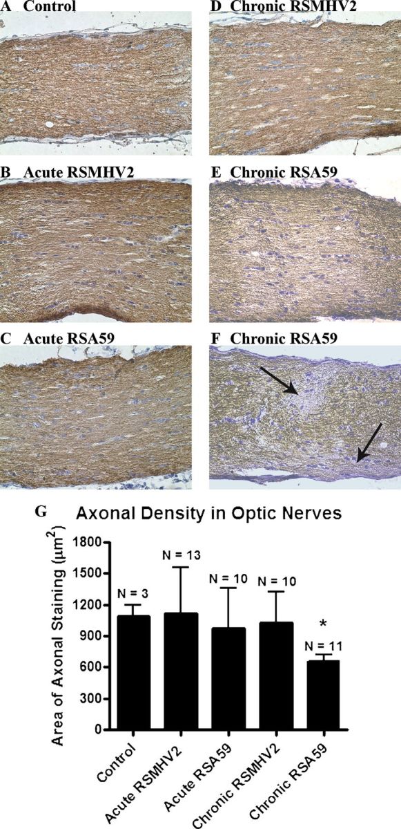
Axon loss in optic nerves of RSA59-infected mice. (A-F) Normal axonal staining is present in a control optic nerve (A), as well as optic nerves from RSMHV2- (B), and RSA5- (C) infected mice at day 6 post inoculation (pi) and at day 30 pi in a RSMHV2-infected mouse (D). Chronic RSA59 infection at 30 days pi resulted in regions of mild diffuse decrease in axonal staining (E) and focal areas with loss of neurofilament staining (arrows) (F) in some mice. (G) The average area of neurofilament-positive axonal staining detected at day 30 pi was significantly lower in optic nerves from RSA59-infected mice than in optic nerves from control, uninfected mice, or RSMHV2-infected mice. *, p = 0.0306. Sections were stained with antineurofilament antibody. Original magnification: (A-F) ×40.