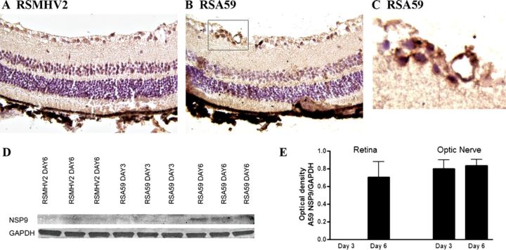
FIGURE 6.
RSA59, but not RSMHV2, spreads from the CNS into the retina. (A-C) No viral antigen staining is present in the retina of an RSMHV2-infected mouse at 6 days post inoculation (pi). Scattered viral antigen-positive cells are found in the retinal ganglion cell layer of the retina of an RSA59-infected mouse (B). Higher magnification view of the boxed area in B shows viral antigen-positive cells (C). (D) Western blot shows the presence of a 13-kb band representing viral antigen in retinal protein extracts from 3 representative RSA59-infected mice at day 6 pi. In contrast, little or no viral antigen is present at day 3 pi with RSA59, and as late as day 6 pi with RSMHV2. (E) The average amount of viral antigen present in protein extracts of optic nerve and retina is shown as the ratio of optical density of MHV nsp9 antibody binding normalized against the binding of glyceraldehyde 3-phosphate dehydrogenase (GAPDH). RSA59 is not detectable in retina until day 6 pi, whereas in optic nerve, it is detected at equally high levels at both days 3 and 6 pi.

