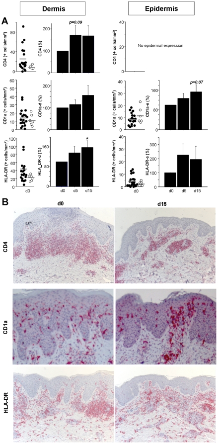
Figure 6. Blue light treatment does not act though similar mechanisms as UV light irradiation.
Skin biopsies of AD patients were obtained before treatment (day 0, d0), on day 5 before irradiation and on day 15 before the last irradiation in the third cycle. Formalin-fixed skin was sectioned and immunohistochemistry performed with anti-CD4, anti-CD1a, and anti-HLA-DR. A, The number of positive cells/mm2 was counted in 5 representative fields per patient both in the epidermis and the dermis. Left panels indicate the mean baseline number for each individual patient (black dots) on d0 with bars showing the mean value of all patients. White dots represent results of healthy control skin from unrelated patients. Right panels contain cumulative data in which the percent change on d5 and d15 to baseline was calculated. Data are shown as mean±SEM (n≥16, * = p≤0.05 as compared to d0). B, Representative stainings for CD4, CD1a and HLA-DR are presented for baseline day 0 and day 15 (x100 magnification for CD4 and HLA-DR, x400 for CD1a).

